Registration for a live webinar on 'Phasing Out Animal Testing: A New Era for Pharmaceutical Innovation' is now open.
See webinar details
This is a limited length demo talk; you may
login or
review methods of
obtaining more access.
- Introduction
-
 63 min
1. Drosophila genetics - the first 25 years
63 min
1. Drosophila genetics - the first 25 years- Prof. Dan Lindsley
- Establishment of the Primary Body Axes
-
 28 min
2. Homeotic genes in Drosophila's bithorax complex - The legacy of Ed Lewis
28 min
2. Homeotic genes in Drosophila's bithorax complex - The legacy of Ed Lewis- Prof. Francois Karch
-
 30 min
- Cell Type Specification and Organ Systems
-
 63 min
4. From germ cell specification to gonad formation
63 min
4. From germ cell specification to gonad formation- Prof. Ruth Lehmann
-
 37 min
5. Drosophila stem cells
37 min
5. Drosophila stem cells- Prof. Michael Buszczak
-
 46 min
6. Legacy of drosophila genetics: female germline stem cells
46 min
6. Legacy of drosophila genetics: female germline stem cells- Prof. Michael Buszczak
-
 34 min
7. Intestinal stem cell-mediated repair in Drosophila 1
34 min
7. Intestinal stem cell-mediated repair in Drosophila 1- Prof. Tony Ip
-
 30 min
8. Intestinal stem cell-mediated repair in Drosophila 2
30 min
8. Intestinal stem cell-mediated repair in Drosophila 2- Prof. Tony Ip
-
 43 min
-
 34 min
10. Axon guidance in Drosophila
34 min
10. Axon guidance in Drosophila- Prof. John Thomas
-
 35 min
11. Development and physiology of the heart
35 min
11. Development and physiology of the heart- Prof. Rolf Bodmer
-
 37 min
12. Identification of host defenses in the Drosophila gut using genome-scale RNAi
37 min
12. Identification of host defenses in the Drosophila gut using genome-scale RNAi- Prof. Dominique Ferrandon
- Genome Organization and Function
-
 53 min
13. The genetic analysis of meiosis in Drosophila melanogaster females
53 min
13. The genetic analysis of meiosis in Drosophila melanogaster females- Prof. R. Scott Hawley
-
 46 min
-
 36 min
15. Dorsal-ventral patterning of the Drosophila embryo
36 min
15. Dorsal-ventral patterning of the Drosophila embryo- Prof. Mike Levine
-
 34 min
-
 27 min
17. Genome-wide pooled CRISPR screen in arthropod cells
27 min
17. Genome-wide pooled CRISPR screen in arthropod cells- Prof. Norbert Perrimon
- Behavior
-
 40 min
-
 54 min
19. Genetics of chemosensory transduction: taste and smell
54 min
19. Genetics of chemosensory transduction: taste and smell- Dr. Leslie Vosshall
-
 20. TRP channels and opsins: universal mediators of sensory biology and behavior
- Prof. Craig Montell
-
 72 min
21. Cracking the case of circadian rhythms by Drosophila genetics
72 min
21. Cracking the case of circadian rhythms by Drosophila genetics- Prof. Jeffrey C. Hall
-
 46 min
22. Sleep in Drosophila
46 min
22. Sleep in Drosophila- Dr. Ralph Greenspan
-
 91 min
-
 34 min
24. Drosophila as a model for drug addiction
34 min
24. Drosophila as a model for drug addiction- Prof. Ulrike Heberlein
- Mechanism of Human Disease
-
 65 min
25. Cross-genomic analysis of human disease genes
65 min
25. Cross-genomic analysis of human disease genes- Prof. Ethan Bier
-
 56 min
26. Human neurodegenerative disease: insights from Drosophila genetics
56 min
26. Human neurodegenerative disease: insights from Drosophila genetics- Prof. Nancy Bonini
-
 21 min
27. Metastasis of Drosophila tumors
21 min
27. Metastasis of Drosophila tumors- Prof. Allen Shearn
-
 51 min
28. Rac-enhanced CAR immunotherapy: RaceCAR
51 min
28. Rac-enhanced CAR immunotherapy: RaceCAR- Prof. Denise Montell
-
 34 min
- Evolution of Adaptive Novelties
-
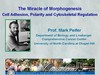
 56 min
30. The evolution of morphological novelty
56 min
30. The evolution of morphological novelty- Prof. Nipam Patel
-
 36 min
31. The genetic architecture of complex traits: lessons from Drosophila
36 min
31. The genetic architecture of complex traits: lessons from Drosophila- Prof. Trudy Mackay
- Archived Lectures *These may not cover the latest advances in the field
-
 25 min
32. Using gene expression information to provide insights into patterning and differentiation
25 min
32. Using gene expression information to provide insights into patterning and differentiation- Prof. Angelike Stathopoulos
-
 49 min
33. Regulation of gastrulation in Drosophila
49 min
33. Regulation of gastrulation in Drosophila- Prof. Dr. Maria Leptin
-
 31 min
34. microRNA function in stem cells
31 min
34. microRNA function in stem cells- Prof. Hannele Ruohola-Baker
Printable Handouts
Navigable Slide Index
- Introduction
- Concept of axon guidance
- Brain section of an adult fly
- Process of path finding by growth cones
- A growth cone
- Embryonic nervous system of a fly
- Navigation of a neuron within the VNC
- Receptors and ligands involved in midline crossing
- Frazzled and Robo function in axon guidance
- Midline crossing: WT versus robo / comm mutants
- Schematic structure of Frazzaled and Robo
- Commissure choice and the Derailed receptor
- RYK subfamily of RTKs
- RYK family of receptors
- Classes of Drl-expressing neurons
- Detection of Drl-expressing neurons
- Drl mutants have defects in midline crossing
- Axon-targeted reporters
- Differential labeling using different reporters
- GAL4 / UAS transactivation
- Expressing Drl in eagle neurons
- Midline crossing in Drl-expressing eagle neurons
- Effect of Drl on non-crossing neurons
- Drl functions as mediator of repulsion
- Wnt5
- Wnt5 expression pattern
- Wnt5 mutants resemble Derailed mutants
- Drl requires Wnt5 to switch axons
- Altering Wnt5 expression
- Repulsive activity of Wnt5 is mediated by Drl
- Soluble Drl-Fc extracellular probe binding in vivo
- Wnt5 binding to Drl in vitro
- Possible signaling of Wnt5 through Fz receptor
- Function of mammalian Wnt protein
- Model of midline crossing
- Integration of the signals in the growth cone
- Major points
Topics Covered
- How axons grow
- The growth cone
- The Drosophila nervous system
- Receptors and ligands involved in axon guidance events
- Attractive and repulsive receptor signaling
- Integration of receptor signals in the growth cone
- Conservation of axon guidance mechanisms during evolution
Links
Series:
Categories:
Therapeutic Areas:
Talk Citation
Thomas, J. (2023, June 19). Axon guidance in Drosophila [Video file]. In The Biomedical & Life Sciences Collection, Henry Stewart Talks. Retrieved April 3, 2025, from https://doi.org/10.69645/FNHL3339.Export Citation (RIS)
Publication History
Financial Disclosures
- Prof. John Thomas has not informed HSTalks of any commercial/financial relationship that it is appropriate to disclose.
Axon guidance in Drosophila

A selection of talks on Neuroscience


Transcript
Please wait while the transcript is being prepared...